Summary
Background/Objective
Obturator hernia is rare type of abdominal hernia and its diagnosis usually is made intraoperatively for bowel obstruction or computed tomography (CT) scans of the abdomen. The aim of this study was to review patients records with respect to clinical manifestation, CT scan findings, and operative outcomes.
Methods
From April 2009 to January 2015, six female patients with incarcerated obturator hernia underwent urgent operation for acute intestinal obstruction. The medical records were reviewed with respect to clinical manifestation, findings of CT scan and the outcomes of operation.
Results
The median age of patients was 83 years (range, 79–87 years) and the body mass index was 21.61 ± 0.52 kg/m2. CT scans of abdomen demonstrated that intestinal obstruction secondary to obturator hernia, consistency with operative findings. Partial bowel resection was performed in two of six patients because of necrosis of incarcerated obturator hernia. The hernia was repaired with interrupted sutures. Lung infection occurred in one patient, and wound infection in another. One recurrence was observed and two patients died from the unrelated diseases during the period of follow-up.
Conclusion
The diagnosis of obturator hernia can be made by CT scan preoperatively, and the obturator hernia should be suspected when an unexplained bowel obstruction in elderly, thin women occurs.
Keywords
bowel obstruction;computed tomography;hernia;laparotomy;obturator
1. Introduction
Obturator hernia is a rare type of hernia in the abdominal wall in which abdominal contents protrude through the obturator canal.1 ; 2 Obturator hernia accounts for 0.07–1% of all hernias and 0.2–1.6% of all cases of mechanical obstruction of the small bowel.3 ; 4 Due to its rarity and unspecific early symptoms, obturator hernia can still be misleading even to the most experienced surgeons. The most common presentation for the patients with obturator hernia is acute intestinal obstruction, and the diagnosis is often made intraoperatively.2; 5 ; 6 As the symptoms are nonspecific and the physical findings are obscure, a correct diagnosis is often delayed until laparotomy for bowel obstruction, despite advances in diagnostic modalities.2 ; 7 It occurs much more in elderly, thin, and multiparous women. Loss of supporting connective tissue and the wider female pelvis are believed to be the cause of obturator hernia.5 ; 8
With the improvement of equipment, a computed tomography (CT) scan is able to detect the obturator hernia preoperatively, being consistent with the findings of laparotomy for obturator hernia.9 ; 10 Early CT imaging allows early diagnosis and reduces morbidity and mortality associated with obturator hernia.10 From April 2009 to January 2015, six consecutive patients with incarcerated obturator hernia underwent laparotomy for acute bowel obstruction in our hospital. The records of the patients were reviewed with respect to clinical manifestation, CT findings, and surgical outcomes.
2. Methods
From April 2009 to January 2015, six patients with incarcerated obturator hernia underwent urgent operation for acute intestinal obstruction in our hospital. Meanwhile, 1950 hernia repairs were done and 728 cases of acute bowel obstruction were admitted to our hospital. Obturator hernia accounts for 0.31% of all hernias and 0.82% of all cases of mechanical obstruction of the small bowel in our hospital. The six patients with incarcerated obturator hernia were female with median age of 83 years (range, 79–87 years). The detailed information of the patents is shown in Table 1. The patients had delivered four children on average and their body mass index was 21.61 ± 0.52 kg/m2. In the emergency room, physical abdominal examination demonstrated that mild abdominal distension and tenderness, especially in the middle and lower abdomen. The patients received abdominal plain x-ray examination and CT preoperatively.
| Age (y) | Parity | BMI (kg/m2) | Bowel resection | Lung infection | Wound infection | Operative time (min) | Hospital stay (d) |
|---|---|---|---|---|---|---|---|
| 83 | 5 | 22.31 | No | No | No | 72 | 9 |
| 87 | 3 | 20.83 | Yes | No | Yes | 106 | 16 |
| 79 | 4 | 21.72 | No | Yes | No | 69 | 10 |
| 84 | 4 | 21.64 | No | No | No | 66 | 8 |
| 81 | 3 | 21.94 | Yes | No | No | 110 | 9 |
| 83 | 6 | 21.23 | no | no | no | 70 | 9 |
BMI = body mass index.
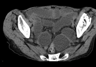
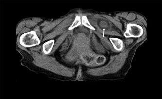
Abdominal X-ray examination showed dilatation of small bowel loops in entire abdomen. CT scan demonstrated dilated bowel loops (Figure 1) and incarcerated bowel between the external obturator and pectineal muscles (Figure 2), suggestive of bowel obstruction secondary to incarcerated obturator hernia. Four of the obturator hernias were in the left and the other two in the right.
|
|
|
Figure 1. Dilated small bowel loops in the lower abdomen and pelvis. |
|
|
|
Figure 2. Incarcerated small bowel between the external obturator and pectineal muscles (arrow). |
All patients underwent urgent laparotomy for acute bowel obstruction. The dilated small bowel occupied the entire abdomen and the herniation of the bowel through the obturator canal was detected, being consistent with the CT scan findings. The incarcerated bowel was reduced gently and carefully inspected. Partial intestinal resection was performed in two patients because of necrosis of incarcerated bowel and no sign of ischemia was noted in the other four patients. The sac was inverted and the defect closed with interrupted sutures. The operative time was 82.17 ± 20.14 minutes, and the length of hospital stay 10.17 ± 2.93 days.
Lung infection occurred in one patient and wound infection developed in another. No hospital death happened in our series. Two patients died from the unrelated diseases during the period of follow-up. One patient developed intermittent abdominal pain, which suddenly appeared and spontaneously disappeared; in this patients, recurrence of obturator hernia was detected by CT scan at 3 years postoperatively. This patient refused further surgical intervention.
3. Discussion
Obturator hernia is a rare type of abdominal hernia with high morbidity and mortality due to delayed diagnosis.2 ; 11 Weakness of the obturator membrane and enlargement of the canal may result in the formation of a hernia sac, which can lead to intestinal incarceration and obstruction. The condition has been nicknamed the little old ladies' hernia as it affects this group due to atrophy and loss of the preperitoneal fat around the obturator vessels in the canal. In our series, the six patients were old and small with median age 83 years and mean body mass index 21.61 ± 0.52 kg/m2.
Incarcerated obturator hernias are usually discovered on abdominal CT scan or emergency surgery due to bowel obstruction.2 ; 7 The usefulness of CT in diagnosis was first reported by Cubillo in 1983 and it has allowed the earlier diagnosis of obturator hernia.9 Nasir et al2 compared the outcomes of obturator hernia between patients who did or did not have a preoperative CT and found that patients with a preoperative CT were less likely to develop a postoperative complication. CT scan is considered the gold standard for diagnosis, whereas ultrasonography and plain radiographs are less specific.
Surgery is the only treatment of choice for obturator hernia. Although surgical repair of obturator hernias has been performed through various approaches, the transabdominal approach is preferred by surgeons as it allows the assessment of the entire abdominal contents and inspection of the incarcerated intestine. We treated the patients using simple suture via laparotomy. In the emergent circumstance of acute intestinal obstruction, laparotomy is a safe and fast way to locate the site of obstruction. The space is limited and dilated intestine is easy to be injured while laparoscopic approach is applied. Therefore, treating the patient with acute bowel obstruction secondary to incarcerated obturator hernia is a challenge to surgeons.
Fast recovery, less postoperative morbidity, and better cosmetic outcomes attract surgeons to employ the laparoscopic technique treating obturator hernia in selected patients.5; 12; 13 ; 14 The trend of surgery for obturator hernia is from open to laparoscopic approach. Ng et al13 reported that there were fewer major complications and less mortality in the laparoscopic group compared with open group in 36 patients undergoing obturator hernia repair during a 15-year period. Regardless of the approach, reduction of the contents and inversion of the hernia sac are the initial steps in the process of operation.
Hernias were repaired by interrupted sutures even in patients without bowel resection in our series. The simple suture was proved to be safe and feasible for incarcerated obturator hernia. We did not use mesh for repair because of the increased risk of the infection. Results from recent studies demonstrated that mesh could be safely applied to obstructed obturator hernia repair.15 ; 16 Mesh repair for the patients with bowel resection is not contraindicated, as long as clean contamination of the wound is maintained during surgery.
In our series, bowel resection and anastomosis were performed in two patients because of the strangulated intestine. Lung infection happened in one patient and wound infection in one patient postoperatively. No hospital death was observed.
In conclusion, obturator hernia is a rare type of hernia, mostly presenting acute intestinal obstruction. The diagnosis of obturator hernia can be made by CT scan preoperatively, and the obturator should be suspected when an unexplained bowel obstruction occurs in elderly, thin women. Mesh repair may have lower long-term recurrence after operation.
References
- 1 Y.H. Ho, H.S. Goh; Obstructed obturator hernia in 90 year olds—a management dilemma; Ann Acad Med Singapore, 20 (1991), pp. 410–411
- 2 B.S. Nasir, B. Zendejas, S.M. Ali, C.B. Groenewald, S.F. Heller, D.R. Farley; Obturator hernia: the Mayo Clinic experience; Hernia, 16 (2012), pp. 315–319
- 3 S.K. Mantoo, K. Mak, T.J. Tan; Obturator hernia: diagnosis and treatment in the modern era; Singapore Med J, 50 (2009), pp. 866–870
- 4 M. Kammori, K. Mafune, T. Hirashima, et al.; Forty-three cases of obturator hernia; Am J Surg, 187 (2004), pp. 549–552
- 5 N.P. Lynch, M.A. Corrigan, D.E. Kearney, E.J. Andrews; Successful laparoscopic management of an incarcerated obturator hernia; J Surg Case Rep, 2013 (2013), p. rjt050
- 6 N. Hodgins, K. Cieplucha, P. Conneally, E. Ghareeeb; Obturator hernia: a case report and review of the literature; Int J Surg Case Rep, 4 (2013), pp. 889–892
- 7 S.W. Gray, J.F. Skandalakis, R.E. Soria, J.S. Rowe Jr.; Strangulated obturator hernia; Surgery, 75 (1974), pp. 20–27
- 8 A.D. Roston, M. Rahmin, A. Eng, A.J. Dannenberg; Strangulated obturator hernia: a rare cause of small bowel obstruction; Am J Gastroenterol, 89 (1994), pp. 277–278
- 9 Y. Yokoyama, A. Yamaguchi, M. Isogai, A. Hori, Y. Kaneoka; Thirty-six cases of obturator hernia: does computed tomography contribute to postoperative outcome?; World J Surg, 23 (1999), pp. 2144–2147
- 10 S.K. Dundamadappa, I.Y. Tsou, J.S. Goh; Clinics in diagnostic imaging; Singapore Med J, 47 (2006), pp. 88–94
- 11 K.V. Chan, C.K. Chan, K.W. Yau, M.T. Cheung; Surgical morbidity and mortality in obturator hernia: a 10-year retrospective risk factor evaluation; Hernia, 18 (2014), pp. 387–392
- 12 Y. Otowa, K. Kanemitsu, Y. Sumi, et al.; Laparoscopic trans-peritoneal hernioplasty (TAPP) is useful for obturator hernias: report of a case; Surg Today, 44 (2014), pp. 2187–2190
- 13 D.C. Ng, K.L. Tung, C.N. Tang, M.K. Li; Fifteen-year experience in managing obturator hernia: from open to laparoscopic approach; Hernia, 18 (2014), pp. 381–386
- 14 T. Yokoyama, A. Kobayashi, T. Kikuchi, K. Hayashi, S. Miyagawa; Transabdominal preperitoneal repair for obturator hernia; World J Surg, 35 (2011), pp. 2323–2327
- 15 T. Karasaki, Y. Nomura, N. Tanaka; Long-term outcomes after obturator hernia repair: retrospective analysis of 80 operations at a single institution; Hernia, 18 (2014), pp. 393–397
- 16 H. Sawayama, K. Kanemitsu, T. Okuma, K. Inoue, K. Yamamoto, H. Baba; Safety of polypropylene mesh for incarcerated groin and obturator hernias: a retrospective study of 110 patients; Hernia, 18 (2014), pp. 399–406
Document information
Published on 26/05/17
Submitted on 26/05/17
Licence: Other
Share this document
Keywords
claim authorship
Are you one of the authors of this document?

